


1
2
3
4
5
6
7
8
9
10
11
12
13
14

Focused Ion Beam Generated cross section imaged using the TEM of Graphene Backgated Field Effect Transistor, Brian Lee, Columbia University, Electrical Engineering
Pseudomonas aeruginosa bacteria, William Cole Cornell, Columbia University, Biological Sciences
Bright Field TEM of Tungsten, Professor Barmak, Columbia University, Applied Physics and Applied Mathematics
Dark Field TEM of Tungsten, Professor Barmak, Columbia University, Applied Physics and Applied Mathematics
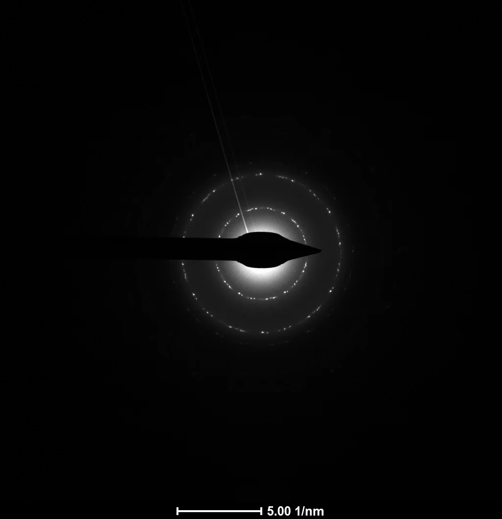
Diffraction pattern of Silicon, Professor Barmak, Columbia University, Applied Physics and Applied Mathematics
High Resolution TEM of Silicon, Professor Barmak, Columbia University, Applied Physics and Applied Mathematics
High Resolution TEM of Silicon, Professor Barmak, Columbia University, Applied Physics and Applied Mathematics
Bright Field TEM of Aluminium, Professor Barmak, Columbia University, Applied Physics and Applied Mathematics
Graphene Diffraction pattern, Professor Barmak, Columbia University, Applied Physics and Applied Mathematics
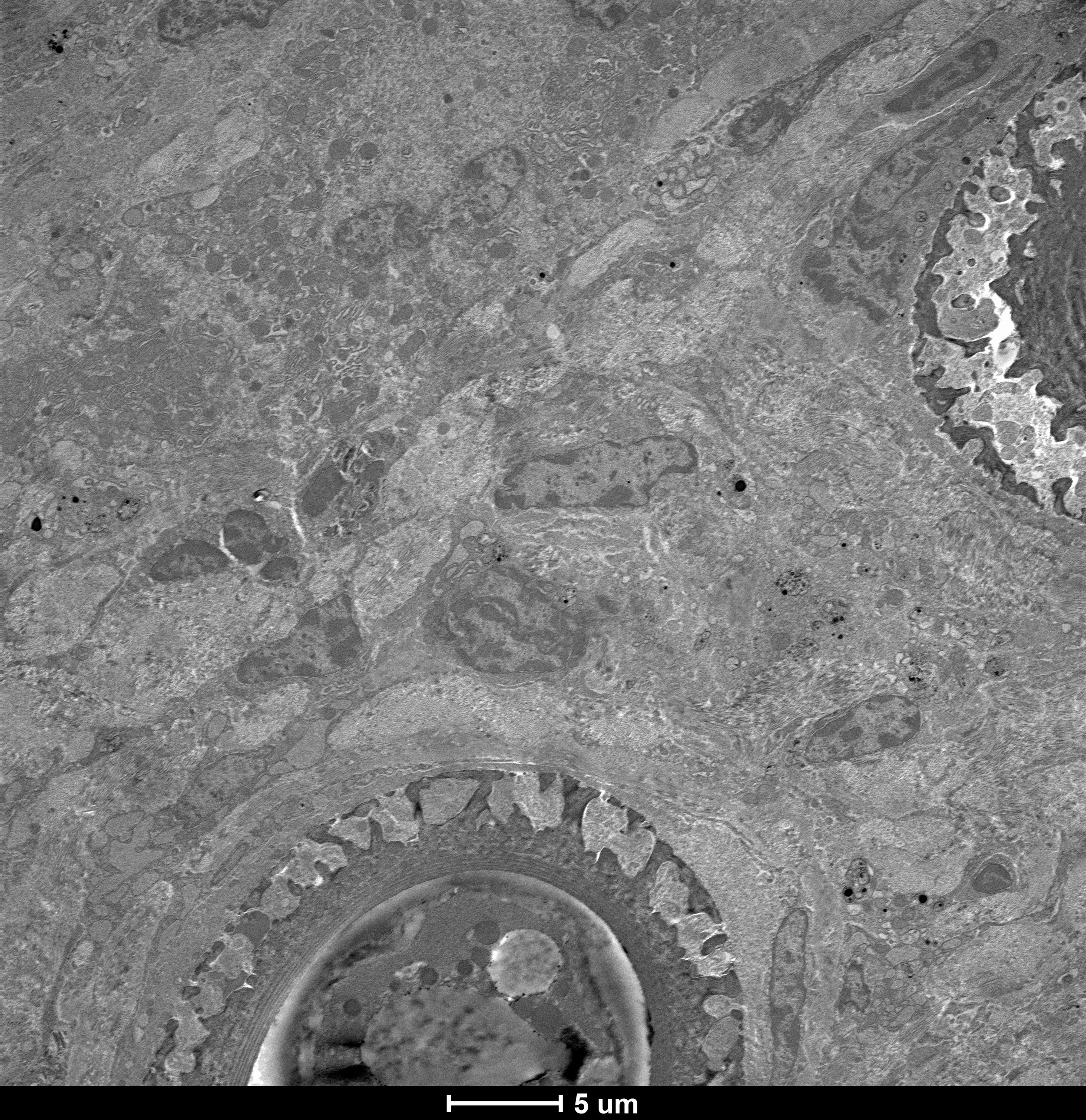
TEM of mouse liver cell showing nucleus, mitochondria, endoplasmic reticulum and golgi apparatus, Simon Williams, Columbia University, Biological Sciences
TEM of mouse liver with nematode infection, Simon Williams, Columbia University, Biological Sciences
Cholesterol crystals in macrophages of a mouse lung, Elizabeth Tarling, University of California Los Angeles, Division of Cardiology
Immunogold labeling of a GFP tagged protein in mouse brain tissue, Chung Dang, Columbia University, Columbia Neuroscience
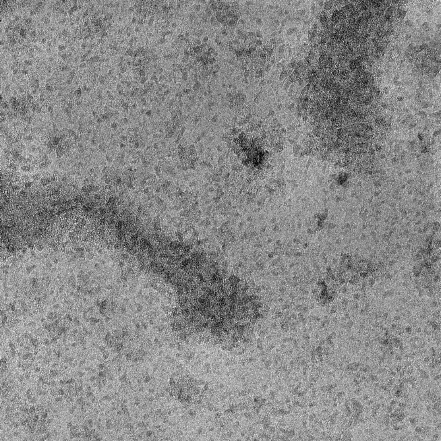
Graphene 80KV Prof. John Kymissis
530 W. 120th St., Schapiro/CEPSR, MC 8903, Suite 1001 New York, NY 10027 / Phone: 212-854-3265
©2016 Columbia University